How the three approaches generally compare
When done well in suitable patients, cancer-control results are often similar. The best approach depends on your tumor, your health and your surgeon’s experience.
Robotic cancer surgery is a minimally invasive, surgeon-controlled operation in which small robotic instruments and a 3D camera, inserted through small incisions, are used to remove a cancerous tumour and, where needed, nearby tissue or lymph nodes.
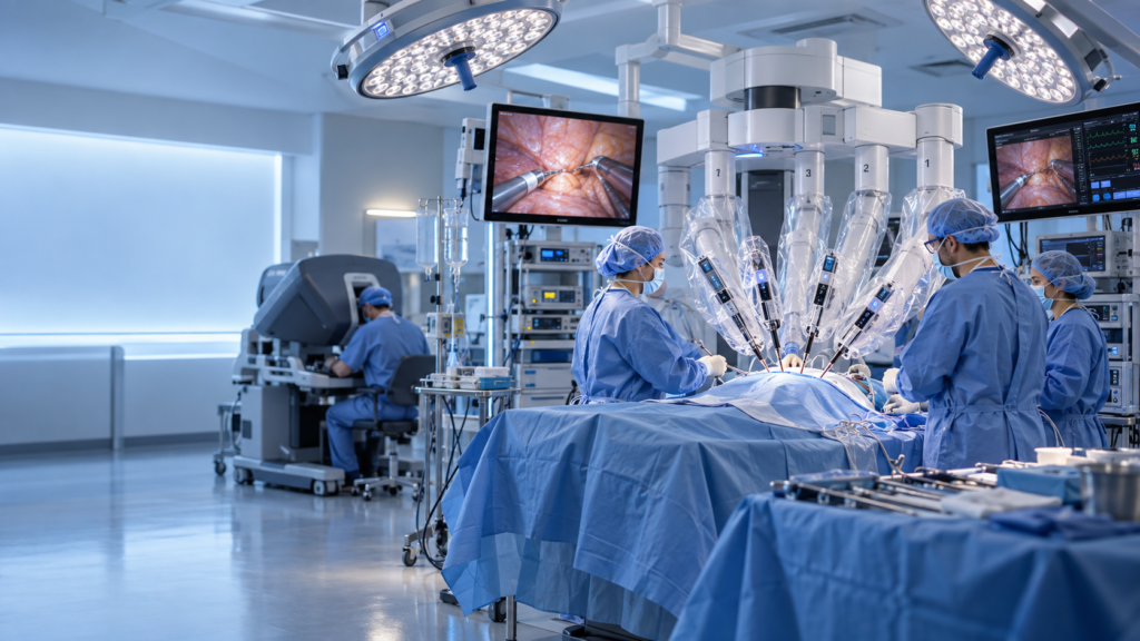
During the procedure, the surgeon sits at a console a few feet from the patient and looks into a magnified 3D high-definition viewer. Hand and finger movements at the console are translated in real time into smaller, steadier movements of the instruments inside the body, while an assistant surgeon and nursing team remain at the bedside.
Although details differ by cancer type and hospital, the operation generally follows these steps:
Robotic surgery is generally considered a safe option when it is performed by an experienced, well-trained surgical team on an appropriately selected patient, with complication rates that are similar to, and for some operations lower than, open surgery.
Like all major surgery, it carries risks. The robotic platform has been used in a very large number of procedures worldwide, but safety for an individual patient depends far more on surgeon experience, hospital volume, patient selection and cancer type than on the robot alone.
Several factors can improve the safety of your operation, including:
Robotic surgery is not automatically better than other approaches; it is a different tool that may reduce pain, blood loss and hospital stay compared with open surgery, while cancer-control results are similar in many well-selected cases.
Compared with conventional laparoscopy, the robot offers 3D vision, wristed instruments and tremor filtration, which can be especially helpful in tight spaces such as the pelvis and chest. The best approach is the one that allows your surgeon to remove the cancer completely and safely.
The table below summarises general differences between the three approaches; individual cases vary:
| Feature | Open Surgery | Laparoscopic | Robotic-Assisted |
|---|---|---|---|
| Incision size | Large (about 10 to 30 cm) | Small (several ports) | Small (several ports) |
| Vision | Direct, naked eye | 2D magnified | 3D magnified, high definition |
| Instrument movement | Hand-held | Straight, limited angles | Wristed, high dexterity |
| Blood loss and pain | Generally higher | Generally lower | Generally lower |
| Typical hospital stay | Longest | Shorter | Shorter |
| Often considered for | Complex or very large tumours | Selected cases | Delicate, deep or confined areas |
Current surgical robots do not operate independently; the surgeon controls every movement of the instruments and remains in the operating room throughout the procedure.
The robot is best described as an advanced surgical tool, not an autonomous machine. It has no decision-making capability and follows the surgeon’s hand movements. Built-in safety systems are designed to pause instrument movement if the surgeon’s head leaves the viewer or a fault is detected.
The da Vinci system, made by Intuitive Surgical, is one of the most widely used surgical robots in the world, and it consists of a surgeon console, a patient-side cart with robotic arms, and a vision system.
Other systems are also available in many countries, including Hugo (Medtronic), Versius (CMR Surgical) and SSI Mantra (developed in India), although availability varies by hospital. Your surgeon’s training and experience with the specific system matter more than the brand name.
The main components of a typical robotic surgery system are outlined below:
| Component | Function |
|---|---|
| Surgeon console | Where the surgeon sits, views the 3D image and controls the instruments |
| Patient-side cart | Robotic arms that hold the camera and wristed instruments |
| Vision cart | Processes and displays high-definition images for the whole team |
Robotic surgery is used to treat many solid tumours in selected patients, particularly those in the pelvis, abdomen, chest and throat, including prostate, kidney, bladder, uterine, colorectal, lung and oropharyngeal cancers.
Suitability depends on tumour size, stage, location and your general health. The table below lists common examples:
| Cancer Type | Common Robotic Operation | Notes |
|---|---|---|
| Prostate | Robotic radical prostatectomy | One of the most common robotic cancer operations |
| Kidney | Partial or radical nephrectomy | Organ-sparing option for suitable small tumours |
| Bladder | Robotic radical cystectomy | Includes urinary diversion; technically demanding |
| Uterine (endometrial) | Robotic hysterectomy and staging | Widely used minimally invasive option |
| Colon and rectum | Robotic colectomy, low anterior resection | May help in a narrow pelvis |
| Lung | Robotic lobectomy or segmentectomy | Early-stage lung cancer |
| Throat (oropharynx) | Transoral robotic surgery (TORS) | Avoids an external neck or jaw incision for some patients |
| Stomach, pancreas, liver, oesophagus | Robotic gastrectomy, pancreatectomy, hepatectomy, oesophagectomy | Selected cases; best done at high-volume centres |
Robotic radical prostatectomy is an established and commonly performed approach for prostate cancer, and in experienced hands, studies report cancer-control outcomes broadly comparable to open surgery.
The 3D magnification and fine instrument control may help the surgeon protect the delicate nerves and muscles that influence erectile function and urinary continence. Outcomes depend on cancer stage and grade, whether nerve-sparing is feasible, your baseline function and, above all, surgeon experience.
If you are considering surgery for prostate cancer, the following points are particularly relevant:
Robotic surgery is a well-established option for uterine (endometrial) cancer, but for early-stage cervical cancer, open radical hysterectomy is now generally recommended because a major randomized trial reported worse survival with minimally invasive surgery.
This is an important example of why cancer type matters when choosing a surgical approach.
The table below summarises the general role of robotic or minimally invasive surgery in gynecologic cancers:
| Cancer | Role of Robotic / Minimally Invasive Surgery |
|---|---|
| Endometrial (uterine) | A standard, well-supported option in suitable patients |
| Cervical (early stage) | Open radical hysterectomy is usually preferred; discuss options carefully with your gynaecologic oncologist, as some very small tumours may have other approaches |
| Ovarian | May be used in selected early-stage cases or for staging; extensive disease often requires open surgery |
Ask your surgeon which approach current guidelines support for your specific stage.
Robotic techniques are used in selected patients with each of these cancers, with potential advantages in confined areas such as the pelvis (rectal cancer), the chest (lung cancer) and the throat (transoral robotic surgery).
How robotic surgery may help varies by cancer type:
The potential benefits of robotic cancer surgery include smaller incisions, less blood loss, reduced pain, a shorter hospital stay and faster recovery compared with open surgery, along with precise tumour removal in skilled hands.
Depending on the operation and your overall health, you may experience some of the following advantages:
These benefits are not guaranteed, and open surgery remains the better choice in some situations.
Robotic cancer surgery carries risks similar to other major operations, including bleeding, infection, anaesthesia reactions, blood clots and injury to nearby organs, along with a small chance of needing to switch to open surgery.
The table below groups the main risks and explains how they are usually managed:
| Risk Category | Examples | How It Is Usually Managed |
|---|---|---|
| General surgical | Bleeding, infection, blood clots | Antibiotics, blood thinners, early walking |
| Anaesthesia related | Nausea, breathing problems, reactions | Pre-operative assessment, anaesthesiologist monitoring |
| Positioning related | Shoulder or nerve strain, facial swelling from head-down position | Careful padding and positioning; often temporary |
| Procedure specific | Urinary leak, bowel leak, lymphocele, sexual dysfunction | Varies by operation; discussed before surgery |
| Conversion | Change to open surgery | Generally a safety decision, not a treatment failure |
Contact your surgeon promptly if you develop fever, worsening pain, heavy bleeding, wound discharge, leg swelling or shortness of breath.
For many well-selected cancers, studies show that robotic surgery achieves cancer-control outcomes comparable to open surgery, provided the tumour is removed completely with clear margins.
The goal is the same for every approach: to remove the entire tumour with healthy margins and to evaluate relevant lymph nodes. Long-term survival depends mainly on cancer stage, tumour biology, complete removal and any additional treatment. Exceptions exist, such as early-stage cervical cancer, where open surgery has been associated with better survival. Ask your surgeon about their own results with your operation, including positive-margin rates.
Many patients experience less pain, less blood loss and smaller scars after robotic surgery than after open surgery, because it avoids a long incision and may reduce tissue handling.
Many patients need fewer strong painkillers and are able to walk within hours of the operation. Small incisions often heal into faint marks over several months. Your own experience will vary with the operation, your anatomy and your overall health.
Robotic surgery may help preserve nerves and organs because magnified 3D vision and precise instruments allow careful dissection around delicate structures, but preservation is attempted only when it is safe for cancer control.
Examples of how robotic techniques may help protect function include:
Cancer control remains the first priority, and preservation is attempted only when it does not compromise complete tumour removal.
You may be a candidate if your tumour can be removed completely through a minimally invasive approach and you are fit for general anaesthesia, but the final decision rests on your imaging, biopsy results and overall health.
The table below contrasts situations that often favour robotic surgery with those where another approach may be safer:
| Often Favourable | May Need Open Surgery or Another Approach |
|---|---|
| Early to locally advanced tumours in suitable locations | Very large tumours, or those invading major blood vessels or organs |
| Good heart and lung fitness | Severe heart or lung disease limiting anaesthesia or head-down positioning |
| Limited prior abdominal surgery | Extensive scarring (adhesions) from previous surgeries |
| Cancers where guidelines support minimally invasive surgery | Cancers where open surgery has shown better survival (e.g., early-stage cervical cancer) |
This decision is usually made with your surgical oncologist, often within a multidisciplinary tumour board.
A well-qualified robotic cancer surgeon is typically certified in the relevant specialty, performs your specific operation frequently and works within a hospital that offers comprehensive cancer and critical care support.
The table below lists what to look for and why each factor matters:
| What to Check | What to Look For | Why It Matters |
|---|---|---|
| Training | Formal robotic training and specialty qualification (surgical oncology, urology, gynaecologic oncology, thoracic or head and neck surgery) | Skill with both the cancer operation and the robotic platform |
| Volume | Regular, high annual number of your exact procedure | Higher experience is associated with better outcomes for many operations |
| Results | Willingness to share complication and margin data | Transparency helps you compare options |
| Team | Multidisciplinary tumour board (surgery, medical and radiation oncology, radiology, pathology) | Treatment is planned as a whole, not surgery alone |
| Facility | Modern robotic system, ICU backup, rehabilitation services | Support if complications occur |
| Openness | Honest discussion of non-robotic alternatives | Confirms the recommendation is based on your needs |
A second opinion is reasonable at any stage, and many surgeons support it.
Ask questions that clarify why robotic surgery is being recommended for you, how experienced your surgeon is, what the alternatives are and what outcomes to expect.
The table below lists key questions and the reason for asking each one:
| Topic | Question to Ask | Why It Matters |
|---|---|---|
| Suitability | Is robotic surgery the best option for my cancer type and stage? | Confirms the approach fits your case |
| Experience | How many of these robotic operations have you performed? | Volume is linked to outcomes |
| Alternatives | What are my other options, including open surgery, radiation or medicines? | Ensures a balanced decision |
| Conversion | What is the chance of conversion to open surgery? | Prepares you for the possibility |
| Results | What are your complication and positive-margin rates? | Helps you judge quality |
| Next steps | Will I need chemotherapy, radiation or other treatment afterward? | Sets expectations for the full plan |
| Side effects | What long-term side effects should I expect? | Supports planning and recovery |
| Cost | What is the estimated total cost and what will insurance cover? | Avoids financial surprises |
Preparation typically involves medical clearance, medication review, nutrition and fitness optimisation, stopping smoking and carefully following your surgical team’s fasting and bowel-preparation instructions.
The table below outlines a typical preparation timeline; your team may adjust it for your situation:
| When | What to Do |
|---|---|
| Weeks before | Complete tests (blood work, ECG, imaging), stop smoking and alcohol, and improve nutrition and activity if advised |
| 1 to 2 weeks before | Review all medicines with your doctor; blood thinners and diabetes medicines often need adjustment |
| Day before | Bowel preparation if required (commonly for colorectal or pelvic surgery); light meals as instructed |
| Day of surgery | Fast as instructed by the anaesthesia team; bring ID, documents and comfortable clothing |
Arrange help at home for the first one to two weeks and discuss any fertility, sexual or emotional concerns with your team before surgery.
Most robotic cancer operations take about 2 to 6 hours, depending on the organ involved, the complexity of the case, the need for lymph node removal and any reconstruction.
You are under general anaesthesia throughout, so you are asleep and unaware of the procedure.
The table below gives general duration estimates; your surgeon can provide a personalised figure:
| Operation | Approximate Duration |
|---|---|
| Robotic prostatectomy | 2 to 4 hours |
| Robotic hysterectomy for cancer | 2 to 4 hours |
| Robotic partial nephrectomy | 2 to 3 hours |
| Robotic colorectal resection | 3 to 6 hours |
| Robotic lobectomy | 2 to 4 hours |
| Robotic cystectomy with diversion | 5 to 8 hours |
Conversion to open surgery is uncommon, but it may be needed if there is unexpected bleeding, dense scarring, difficult anatomy or a tumour that cannot be removed safely with the robotic approach.
Consent for robotic surgery typically includes consent for possible conversion. Conversion is generally regarded as a safety decision rather than a treatment failure. Ask your surgeon for their conversion rate for your specific operation.
Many patients go home within 1 to 5 days after robotic cancer surgery and return to light daily activities within 2 to 4 weeks, with full recovery typically taking 4 to 8 weeks, depending on the operation.
The table below shows typical timelines by operation; your own recovery may be faster or slower:
| Operation | Typical Hospital Stay | Return to Desk Work |
|---|---|---|
| Prostatectomy | 1 to 2 days | 2 to 4 weeks |
| Hysterectomy | 0 to 1 day | 2 to 4 weeks |
| Kidney (partial nephrectomy) | 1 to 3 days | 2 to 4 weeks |
| Colorectal resection | 3 to 5 days | 3 to 6 weeks |
| Lung lobectomy | 2 to 4 days | 3 to 6 weeks |
Age, fitness, other medical conditions and any complications can all affect these timelines.
Most surgeons advise walking daily, avoiding heavy lifting for roughly 4 to 6 weeks, taking prescribed medicines and keeping incisions clean and dry, but your own instructions may differ, so follow your surgical team’s guidance.
The table below summarises common aftercare guidance:
| Area | General Guidance | Note |
|---|---|---|
| Walking | Short walks starting the day of surgery or as advised | Helps reduce the risk of blood clots |
| Lifting | Avoid lifting more than about 4 to 5 kg (10 lb) and strenuous exercise until cleared | Usually for about 4 to 6 weeks |
| Nutrition | Balanced, protein-rich meals and adequate fluids unless restricted | Supports healing |
| Driving | Wait until you are off opioid pain medicines and can move comfortably | Confirm with your surgeon |
| Wound care | Keep incisions clean and dry as instructed | Ask about showering and dressings |
| Follow-up | Attend all scheduled visits, tests and scans | Needed to monitor recovery and cancer status |
Certain symptoms need prompt medical attention. The table below explains what to watch for and what to do:
| Warning Sign | Suggested Action |
|---|---|
| Fever of 38 °C (100.4 °F) or higher | Contact your surgeon or hospital the same day |
| Severe or worsening pain | Contact your surgical team promptly |
| Heavy bleeding or foul wound discharge | Contact your surgical team promptly |
| Calf pain or leg swelling | Seek urgent medical care (possible blood clot) |
| Chest pain or shortness of breath | Seek emergency care immediately |
Some patients need additional treatment after robotic surgery, and that decision depends mainly on pathology results, cancer stage, margin status and lymph node findings rather than on the surgical approach.
After surgery, the removed tissue is examined by a pathologist, and your tumour board then recommends the next step.
The table below shows how pathology findings may guide further treatment:
| Pathology Finding | Possible Next Step |
|---|---|
| Cancer completely removed, early stage | Observation with regular follow-up |
| Positive or close margins | Additional surgery or radiation |
| Lymph node involvement | Chemotherapy, radiation, hormone or targeted therapy |
| High-risk features | Adjuvant systemic therapy or immunotherapy where appropriate |
Robotic cancer surgery often costs more than open or conventional laparoscopic surgery because of equipment and instrument costs, although a shorter hospital stay may offset part of the difference.
Total cost varies widely by country, hospital, room category, surgeon, cancer type and the supplies required, and insurance coverage varies as well.
The table below lists the main factors that influence the final bill:
| Cost Factor | How It Affects the Bill |
|---|---|
| Type and complexity of operation | Longer, more complex operations cost more |
| Hospital category and city | Facility charges differ widely |
| Length of stay and ICU need | Each additional day adds room and nursing charges |
| Robotic instruments and consumables | Single-use and limited-use instruments add cost |
| Tests and follow-up | Pathology, imaging and post-operative care are often billed separately |
To manage costs and avoid surprises, follow the steps below in order:
| Step | Action | Who to Contact |
|---|---|---|
| 1 | Request a written, itemized estimate | Hospital billing or patient services |
| 2 | Ask whether robotic surgery is covered and whether any sub-limits apply | Insurer or TPA |
| 3 | Obtain pre-authorization before the procedure | Hospital insurance desk with your insurer |
| 4 | Check eligibility for employer, corporate or government health schemes | Employer HR or scheme office |
Many insurers cover robotic surgery when it is medically necessary, but policy terms differ, so confirm details before you proceed.
The future of robotic cancer surgery is expected to include more accessible robotic platforms, better imaging, AI-assisted planning and improved training, all aimed at making cancer surgery more precise and less invasive.
Several developments are being researched or introduced, including:
Even as technology advances, the surgeon’s skill, judgment and a multidisciplinary team are expected to remain the most important factors in your outcome.
Robotic cancer surgery is available at several major hospitals in Bengaluru (Bangalore), a city regarded as a medical hub in South India, although the robotic systems, the cancers treated and surgeon experience differ from one hospital to another.
Patients from Karnataka, other Indian states and other countries travel to Bengaluru for advanced cancer care. The most useful question is not whether a hospital owns a robot, but whether it has a surgeon who regularly performs your specific operation within a full cancer-care team.
Because hospital experience matters more than equipment alone, it helps to raise a few points when you first enquire. Consider asking about the following:
The table below shows the types of robotic cancer surgery commonly offered by large cancer-care centers and what to confirm with your chosen hospital in Bangalore:
| Cancer Area | Robotic Operations Often Offered | Confirm With the Hospital |
|---|---|---|
| Urologic (prostate, kidney, bladder) | Prostatectomy, partial or radical nephrectomy, cystectomy | Surgeon volume for your operation; urinary diversion experience |
| Colorectal | Colectomy, low anterior resection, abdominoperineal resection | Rectal cancer volume; stoma care support |
| Upper gastrointestinal (esophagus, stomach, pancreas, liver) | Esophagectomy, gastrectomy, selected pancreatic and liver operations | Team experience with these complex operations; ICU support |
| Gynecologic | Hysterectomy, staging, lymph node procedures | Guideline-based approach for cervical cancer |
| Thoracic (lung, thymus) | Lobectomy, segmentectomy, thymectomy | Thoracic robotic experience; pulmonary rehabilitation |
| Head and neck | Transoral robotic surgery (TORS) | Whether TORS is offered; speech and swallowing therapy |
Services change over time, so confirm current systems, surgeons and availability directly with the hospital before planning treatment.
A sound choice in Bangalore combines a qualified surgeon with regular experience in your operation, a hospital with recognized accreditation and a multidisciplinary tumor board, and clear communication about outcomes, alternatives and costs.
The table below lists checks worth making before you decide:
| Check | What to Ask | Why It Matters |
|---|---|---|
| Accreditation | Is the hospital accredited (for example NABH or JCI)? | Indicates adherence to recognized quality and safety standards |
| Surgeon credentials | Is the surgeon registered with the medical council and qualified in the relevant cancer specialty? | Confirms formal training |
| Case volume | How many of my specific operation are performed here each year? | Experience is linked to outcomes for many operations |
| Tumor board | Will my case be reviewed by a multidisciplinary team? | Treatment is planned as a whole, not surgery alone |
| Support services | Are dietitian, physiotherapy, stoma care and supportive care available? | Recovery depends on more than the operation |
| Critical care | Are ICU, blood bank and emergency backup on site? | Support if complications occur |
| Communication | Are consultations available in a language I am comfortable with? | Clear understanding supports informed consent |
A second opinion is reasonable at any stage, and scans and reports can often be shared for review.
The cost of robotic cancer surgery in Bangalore varies by hospital, cancer type, room category and length of stay, so a helpful first step is to request a written, itemized estimate in Indian rupees (INR) that states clearly what is included and excluded.
Package prices can look similar but cover different things. Comparing like with like helps you avoid surprises.
The table below shows what to confirm in any estimate:
| Item | What to Confirm |
|---|---|
| Surgeon and anesthesia fees | Whether they are included in the package |
| Robotic instruments and consumables | Whether single-use items are included |
| Room category and ICU | Number of days covered and daily rates beyond that |
| Tests, pathology and imaging | Which are included before, during and after surgery |
| Medicines and supplies | What is included at discharge |
| Complications or extended stay | How additional charges are handled |
If you are travelling from another city or country, the table below outlines common planning points:
| Planning Item | General Guidance |
|---|---|
| Before travel | Share reports and scans for a teleconsultation, and carry biopsy reports and slides or blocks |
| Before surgery | Arrive a few days early for tests and anesthesia review, as advised |
| After discharge | Many patients stay nearby for about 1 to 2 weeks for check-ups, depending on the operation |
| Air travel | Travel only after your surgeon confirms it is safe; the timing varies by operation |
| International patients | A medical visa may be required; check current rules with the embassy or consulate and the hospital’s international patient desk |
| Insurance | Ask about cashless approval, pre-authorization and acceptance of your insurer |
| Follow-up | Agree in advance on teleconsultation and how a local doctor will be involved |
Robotic surgery has important limitations: it has not been shown to improve cure rates over well-performed laparoscopic or open surgery for many cancers, it typically requires specialized training, and it may take longer, cost more and be unavailable at some hospitals.
Understanding these limits helps you weigh the technology realistically. The robot is a tool that can support a skilled surgeon, but it does not replace surgical judgment or experience.
The table below summarizes the main limitations and what each means for you:
| Limitation | What It Means for the Patient |
|---|---|
| Cure-rate advantage not demonstrated | For many cancers, cancer-control results are often similar to good laparoscopic or open surgery; the benefit is usually less physical strain, not a higher cure rate |
| Learning curve | Outcomes depend on the surgeon and team; less experienced teams may have longer operations and higher complication rates |
| Limited touch feedback | Many systems give little or no sense of touch, so surgeons rely mainly on vision |
| Setup and operating time | Docking the robot can add time; some operations take longer than open surgery |
| Higher cost | Equipment and single-use instruments often raise the bill, and coverage varies |
| Not suitable for every case | Very large, advanced or emergency cases may need open surgery |
| Availability | Not every hospital or city has a robotic system or a surgeon experienced in your operation |
Patients who may benefit most are those whose tumor lies in a deep, narrow or hard-to-reach area, who need delicate dissection or reconstruction, or who are likely to tolerate a large open incision poorly, provided the cancer itself is suitable for a minimally invasive approach.
The table below outlines common situations in which robotic surgery may offer an advantage, and the reason:
| Patient or Tumor Situation | Why Robotic Surgery May Help |
|---|---|
| Narrow pelvis (for example rectal or prostate cancer) | 3D vision and wristed instruments can make work in tight spaces easier |
| Need for nerve preservation | Fine, steady movements may help protect nerves that affect urinary and sexual function |
| Higher body weight | Access may be easier than with straight laparoscopic instruments in some patients |
| Complex suturing or reconstruction (for example partial nephrectomy, bladder reconstruction) | Wristed instruments can make precise stitching more manageable |
| Older or less fit patients who can safely undergo anesthesia | Smaller incisions may reduce wound-related strain and speed early walking |
| Early-stage cancers where minimally invasive surgery is standard (for example prostate, endometrial, kidney) | Robotic surgery is a well-established option in experienced hands |
Whether you personally benefit depends on your cancer, your anatomy and your surgeon’s experience. To understand the reasoning for your case, consider asking:
Robotic surgery may offer little or no advantage, or may be unsuitable, for patients with very large or locally advanced tumors, extensive previous surgery, severe heart or lung disease, emergency conditions, and cancers where open surgery has shown better survival, such as early-stage cervical cancer.
The table below lists situations in which another approach is often considered, along with the usual reason:
| Situation | Reason for Caution | Approach Often Considered |
|---|---|---|
| Very large tumors or tumors invading nearby organs or vessels | Safe and complete removal may be difficult | Open surgery, or combined treatment first |
| Extensive prior abdominal or pelvic surgery | Dense scarring (adhesions) can make access risky | Open or hybrid approach |
| Severe heart or lung disease | Gas inflation and head-down positioning may strain the body | Alternative anesthesia plan, open surgery or non-surgical treatment |
| Emergency situations (for example bowel obstruction or perforation) | Speed and flexibility are priorities | Open or laparoscopic surgery |
| Early-stage cervical cancer (many cases) | A randomized trial reported worse survival with minimally invasive radical hysterectomy | Open radical hysterectomy |
| No access to an experienced robotic team | Results depend heavily on surgeon experience | Referral to an experienced center or an alternative approach |
These are general patterns, not fixed rules. Your surgical oncologist will review your scans, pathology and health before recommending an approach.
Robotic surgery is an established option for suitable rectal cancers, where 3D vision and wristed instruments can help the surgeon work in the narrow pelvis, and studies report cancer-control outcomes broadly comparable to laparoscopic surgery, although evidence of clear superiority is mixed.
The central principle of rectal cancer surgery is total mesorectal excision (TME), which removes the rectum together with its surrounding fatty tissue and lymph nodes. Many patients with locally advanced rectal cancer receive chemotherapy, radiation or both before surgery, and MRI helps plan the operation.
The table below lists the main robotic rectal operations and when each is typically used:
| Operation | Typically Used For | Key Point |
|---|---|---|
| Low anterior resection (LAR) | Upper and mid rectal tumors | Sphincter preserved; a new join (anastomosis) is made |
| Ultra-low or intersphincteric resection | Low tumors near the anal canal | Aims to preserve continence; function varies |
| Abdominoperineal resection (APR) | Very low tumors involving the sphincter muscles | A permanent colostomy is generally needed |
Once the type of operation is chosen, patients usually have practical questions about function and recovery. The points below cover concerns that are commonly raised:
A temporary stoma is often created after low rectal resections to protect the new join while it heals, whereas a permanent stoma is generally needed only when the anal sphincter muscles cannot be preserved safely.
The table below shows how the operation type relates to the likelihood of a stoma:
| Situation | Stoma Outcome | Typical Reversal Timing |
|---|---|---|
| Low anterior resection with a low join | A temporary loop ileostomy is common | Often about 2 to 6 months later, and later if chemotherapy is needed |
| High rectal resection with a safe join | A stoma may not be needed | Not applicable |
| Abdominoperineal resection | Permanent colostomy | Generally permanent |
Even when no permanent stoma is needed, the rectum’s role in storing stool is altered, so bowel habits often change for a period. Common experiences include:
Together these symptoms are called low anterior resection syndrome (LARS). A stoma nurse, dietitian and pelvic floor physiotherapist can help.
Robotic colectomy is a suitable option for many colon cancers, but for straightforward cases laparoscopic surgery gives broadly similar results, so the robot’s advantages are more likely in technically complex operations.
Robotic surgery may make it easier to join the bowel ends inside the body and to work precisely around blood vessels, although operating time and cost are often higher. Emergency operations for obstruction or perforation are usually performed by open or laparoscopic methods.
The table below lists common robotic colon operations by tumor location:
| Operation | Tumor Location | Note |
|---|---|---|
| Right hemicolectomy | Cecum, ascending colon, hepatic flexure | Often with complete mesocolic excision; the join may be made inside the body |
| Transverse colectomy | Transverse colon | Less common; technically variable |
| Left hemicolectomy | Descending colon | Requires careful vessel and nerve handling |
| Sigmoid colectomy | Sigmoid colon | Commonly performed robotically |
| Subtotal or total colectomy | Multiple tumors or inherited conditions (for example Lynch syndrome) | Selected cases |
Many patients having robotic colon cancer surgery follow an enhanced recovery (ERAS) pathway, which typically means early walking, early eating and a hospital stay of about 3 to 5 days, although bowel function and energy return at different rates.
The table below shows typical milestones; your own timeline may be faster or slower:
| Milestone | Typical Timing |
|---|---|
| Start walking | Same day or the next day |
| Drinking and light diet | Within 1 to 2 days |
| Passing gas and stool | Within a few days |
| Hospital discharge | About 3 to 5 days |
| Pathology report | About 1 to 2 weeks |
| Chemotherapy, if advised | Often starts about 4 to 8 weeks after surgery |
| Return to desk work | About 3 to 6 weeks |
Contact your surgical team promptly if you develop fever, worsening abdominal pain, repeated vomiting, a swollen abdomen, or inability to pass gas or stool.
Robotic esophagectomy is an option at experienced centers for selected patients with esophageal cancer, and it aims to reduce the physical impact of a very major operation, but results depend heavily on team experience and often on treatment given before surgery.
Many patients receive chemotherapy or chemoradiation before surgery, and the operation is often planned several weeks afterward. Early randomized data suggest robotic-assisted approaches may reduce some lung complications compared with open surgery, but the evidence is still developing.
The table below outlines the main types of esophagectomy, which may be done robotically in part or in full:
| Operation | Approach | Where the New Join Is Made |
|---|---|---|
| Ivor Lewis esophagectomy | Abdomen and right chest | In the chest |
| McKeown (three-field) esophagectomy | Abdomen, chest and neck | In the neck |
| Transhiatal esophagectomy | Abdomen and neck | In the neck |
Because esophagectomy is a major operation, careful preparation supports both safety and results. Before recommending surgery, your team typically completes the following steps:
Esophagectomy carries higher risks than many other cancer operations, including leakage at the new join, pneumonia and swallowing problems, and recovery is longer, with a hospital stay of roughly 1 to 2 weeks and a gradual return to eating over weeks to months.
The table below lists the main concerns and how they are usually managed:
| Concern | What It Means | How It Is Usually Managed |
|---|---|---|
| Leak at the join (anastomotic leak) | Leakage where the esophagus is reconnected | Swallow tests, drainage, antibiotics, endoscopic stents or further surgery |
| Pneumonia and breathing problems | Chest infection after major surgery | Breathing exercises, early walking, physiotherapy |
| Narrowing (stricture) | Scar tightening at the join, causing swallowing difficulty | Endoscopic dilation |
| Reflux and regurgitation | Stomach contents rising after surgery | Small meals, sleeping with the head raised, medicines |
| Weight loss and malnutrition | Common after esophagectomy | Dietitian support and sometimes a temporary feeding tube |
Eating usually restarts in stages, beginning with liquids and progressing to soft foods, following your team’s plan.
Robotic gastrectomy is used for selected stomach cancers without extensive spread, and studies report short-term outcomes similar to laparoscopic surgery, although long-term evidence is still developing and thorough lymph node removal is central to cancer control.
Many patients with locally advanced stomach cancer receive chemotherapy before and after surgery. Robotic techniques may help with precise lymph node dissection and reconstruction, and they are often best performed at high-volume centers.
The table below outlines the main types of gastrectomy and how the digestive tract is rebuilt:
| Operation | Used When | Reconstruction |
|---|---|---|
| Distal (subtotal) gastrectomy | Tumors in the lower stomach | Roux-en-Y or Billroth-type connection |
| Total gastrectomy | Tumors in the upper or middle stomach or spreading widely in the stomach wall | Esophagus joined to the small bowel (Roux-en-Y) |
| Proximal gastrectomy | Selected early tumors in the upper stomach | Reconstruction designed to limit reflux |
After a gastrectomy many patients need to eat smaller, more frequent meals and may experience early fullness, dumping symptoms and weight loss, but a dietitian and a staged diet plan can help you adapt.
The table below summarizes common changes and practical tips:
| Change | What to Expect | Practical Tip |
|---|---|---|
| Meal size | Smaller stomach capacity and early fullness | Eat about 5 to 6 small meals a day |
| Dumping syndrome | Cramps, sweating, dizziness or diarrhea after meals | Limit sugary foods; take fluids apart from meals |
| Weight loss | Common in the first months | Choose protein-rich foods; review with a dietitian |
| Vitamin and mineral needs | Vitamin B12, iron and calcium may fall, especially after total gastrectomy | Take supplements or injections as advised |
| Diet progression | Liquids, then soft foods, then regular textures over weeks | Follow your team’s plan |
Robotic thoracic surgery is commonly used for early-stage lung cancer and for selected tumors of the mediastinum, such as thymoma, through small chest incisions.
The table below lists common robotic thoracic operations:
| Condition | Robotic Operation | Notes |
|---|---|---|
| Early-stage lung cancer | Lobectomy or segmentectomy with lymph node removal | Depends on tumor size, location and lung function |
| Thymoma and thymic tumors | Thymectomy | Suitable for selected tumors that have not invaded nearby structures |
| Mediastinal masses | Removal of selected cysts or nerve-related tumors | Depends on size and location |
| Esophageal cancer | Robotic esophagectomy | Covered in the esophageal cancer questions above |
Lung function tests, imaging and stopping smoking are important parts of preparation.
Robotic surgery and video-assisted thoracoscopic surgery (VATS) both offer minimally invasive lung cancer surgery with comparable cancer outcomes in many studies, and the choice usually depends on tumor factors, surgeon experience and availability.
The robot may provide 3D vision and greater instrument dexterity, which can help in complex resections, whereas VATS is well established, widely available and often less costly.
The table below compares the two approaches in general terms:
| Feature | VATS | Robotic |
|---|---|---|
| Instruments | Straight, rigid | Wristed, greater dexterity |
| Vision | 2D or 3D depending on system | 3D, high definition |
| Availability | Available in many centers | Limited to hospitals with a robotic system |
| Cost | Generally lower | Generally higher |
| Cancer outcomes | Comparable in many studies | Comparable in many studies |
Recovery after either approach follows a broadly similar pattern. Points that patients commonly ask about include:
Robotic hysterectomy with removal of the fallopian tubes and ovaries, along with lymph node assessment where indicated, is a well-established minimally invasive option for many endometrial cancers, and it may be especially helpful for patients with higher body weight.
The table below outlines the main steps of a typical robotic operation for endometrial cancer:
| Step | What Is Done |
|---|---|
| Hysterectomy | The uterus and cervix are removed |
| Salpingo-oophorectomy | The fallopian tubes and ovaries are usually removed |
| Lymph node assessment | Sentinel lymph node mapping with fluorescent dye, or lymph node removal, depending on risk |
| Specimen removal | The uterus is removed in a protective bag, generally through the vagina, to reduce the risk of spread |
| Laboratory testing | Pathology and molecular testing guide the need for further treatment |
Hospital stay is often 0 to 1 day. If both ovaries are removed before menopause, menopausal symptoms may begin, which your team can help manage.
Fertility preservation is possible only in carefully selected early-stage cases, and the options depend on the cancer type, so it should be discussed with a gynecologic oncologist and a fertility specialist before treatment begins.
The table below outlines general possibilities by cancer type:
| Cancer | Possible Fertility-Sparing Approach | Important Note |
|---|---|---|
| Early endometrial (low grade, very early stage) | Hormonal therapy with close monitoring in carefully selected patients | Requires strict follow-up; hysterectomy is often advised after childbearing |
| Early cervical (small, low-risk tumors) | Less extensive surgery such as cone biopsy or trachelectomy in selected cases | The surgical route is chosen carefully because a randomized trial reported worse survival with some minimally invasive radical operations |
| Early ovarian (selected types) | Removal of one ovary and tube with staging in selected patients | Depends on tumor type and grade |
Egg or embryo freezing may be an option before treatment, so raise fertility early in your consultation.
If you are considering robotic cancer surgery, book a consultation with a qualified surgical oncologist to review your scans, pathology and all treatment options, including a second opinion if you wish. You may also find Preparing for Cancer Surgery and Questions Patients Really Ask helpful.